
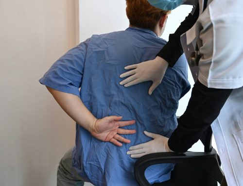
80% of people experience some form of general back pain at some point in their life, with 90% of that pain being specifically low back pain. The most common form of back pain is designated nonspecific or mechanical because it lacks a specific pathology that has caused the pain. The good news is that nonspecific back pain cases are typically self-limiting, resolving in 4-6 weeks. Although low back pain is common, it can greatly affect our day to day life preventing us from playing sports, doing regular work, affecting our sleep or preventing us from going on regular walks. When people go to their doctor for low back pain, they have many questions with one of the more common ones being, “do I need an x-ray or MRI?” A valid question that we will answer below.
Shoulder I get Imaging?
In 2011, Chou et al published a summary on the evidence for routine imaging of low back pain. They found that the American College of Physicians Low Back Pain Guideline recommends selective imaging for patients in whom it is clinically indicated. Furthermore, a meta-analysis of 6 random control trials found no difference between routine low back imaging and usual care without imaging in terms of pain, function, quality of life, or overall patient-reported improvement. In other words, imaging can be accurate at identifying conditions but it does not improve outcomes or affect the treatment plan. For short-term outcomes (less than 3 months), trends actually favoured usual care without imaging. But why is that?
Why Do I Not Need Imaging for my Low Back Pain?
Here are some of the main reasons imaging is not typically needed for low back pain.
1. Asymptomatic Findings
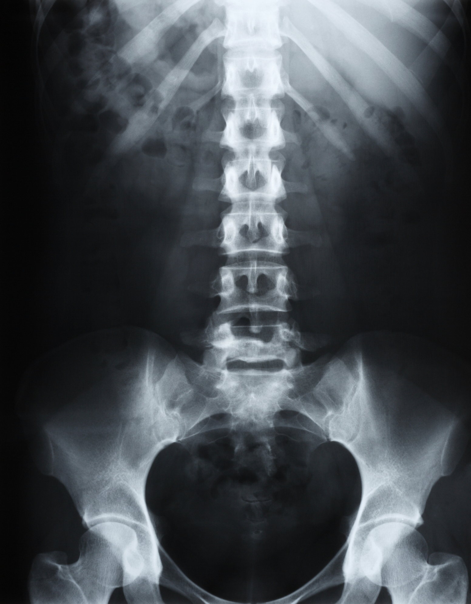
Most lumbar imaging abnormalities are common in people without low back pain and are loosely associated with symptoms. They are so commonly found in advanced imaging of asymptomatic persons that they could be interpreted as normal signs of aging. 36% of asymptomatic people over the age of 60 years old had herniated discs, 21% had spinal stenosis, and more than 90% had degenerated or bulging discs. Remember that low back pain has a favourable natural history with most people experiencing substantial improvement over the next 4 weeks. So imaging may lead to unnecessary procedures or increased anxiety in patients.
It can be devastating to hear that you have “degenerative disc disease” or some other incidental finding on imaging of your low back, potentially impacting your recovery in a negative way. But it is important to remember that these findings can be incidental, may not be the cause of your back pain and can be considered completely normal. A good analogy is grey hair. Like disc degeneration, some people get grey hair earlier than others, but it is a normal process of aging, and not necessarily a cause of major concern.
2. Low Specificity
When considering CT scans, it is important to note that they have a 30% false positive rate. A false positive is when a test indicates that you have particular diagnosis when, in reality, it is not true. This happens 30% of the time with CT scans. In regard to MRIs, they have are highly sensitive but have a 20% specificity for a given condition. A test with a low specificity is more likely to produce a false positive rate. This is not to say that CT scans and MRIs are pointless tools, but rather that they should only be used when necessary instead of routinely.
3. Major Pathologies
When we have pain, we often get anxious and our mind focuses on the worst case scenario. So patients often wish to get imaging done to hopefully calm their mind. Rest assured, clinicians are well trained to pick up on more sever pathologies such as cancer, infection, and cauda equina syndrome. These pathologies have identifiable risk factors that are investigated during the history, making routine imaging redundant and unnecessary.
When to Get Imaging
Although we have talked at length why imaging is not always routinely used to diagnose patients with low back pain, it is important to mention the instances when they shoulder be considered.
- Back pain after sever trauma (fall >10 feet, car accident over 80 km/h, concurrent neck fracture, midline spine tenderness, progressive neurological deficits, altered consciousness)
- Suspected degenerative spondylolisthesis or stenosis with progressive neurological deficits in patients >50 years old
- Potential or serious pathology suspected from the history or physical exam (suspected cancer, inflammatory arthritis)
- After failed conservative therapy (4-6 weeks)
- Adult patient with progressive scoliosis
There can be various reasons why a practitioner may not follow the above guidelines and request imaging when it is not necessary. Potential reasons can include: lack of knowledge of the guidelines, fear of missing a diagnosis, pressure from the patient or that there is no other treatment options.
In summary, routine imaging of the lumbar spine for non-complicated low back pain is not commonly needed and can cause unnecessary fear and worris that might actually cause an increase in back pain disability. Findings on imaging rarely change the treatment plan and course of action to get the patient’s pain experience resolved. In most cases, it is worth a 4-8 week trial of care.
Check out of YouTube video on this topic by clicking HERE!
Looking to learn more about low back pain? Click HERE to read another one of our blog posts on patterns of low back pain.
Want to book an appointment with one of our chiropractors or massage therapist? Click HERE to get started.
REFERENCE
Chou et al. Diagnostic imaging for low back pain: advice for high-value health care from the American College of Physicians. Ann Intern Med. 2011. 154(3):181-189. Available from: https://pubmed.ncbi.nlm.nih.gov/21282698/

