
One of the most common questions I hear from patients with low back pain is:
“Should I get an X-ray or MRI to see what’s going on?”
Most of the time, the answer is no — at least not initially.
That can seem surprising. If your back hurts, particularly if the pain is severe, it seems logical that taking a picture of your spine would help us find the problem.
But one of the things I often tell patients is:
Imaging can take a picture of you, but it can't necessarily tell me how you're feeling.
For most uncomplicated episodes of low back pain, clinical guidelines recommend against routine imaging. An X-ray, CT scan or MRI usually doesn't improve recovery or change the initial treatment plan when there are no signs of a serious underlying condition.
That doesn't mean imaging isn't useful. It can be extremely important when we have a specific clinical question that the image can help answer.
The important question isn't simply, “Can we image your back?”
It's “Will imaging provide information that changes what we do next?”
Why Isn't Imaging Routinely Recommended for Low Back Pain?
Research comparing immediate imaging with usual clinical care has found that routine imaging in people without signs of serious underlying disease does not improve pain, function, quality of life or overall recovery.
This is why guidelines from organizations including the American College of Radiology and Choosing Wisely Canada recommend a selective approach to imaging.
There are several reasons.
1. Your MRI Doesn't Necessarily Explain Your Pain
One of the challenges with spinal imaging is that abnormalities are incredibly common — including in people who have no back pain whatsoever.
A large systematic review looked at CT and MRI findings in more than 3,000 people without symptoms. The prevalence of degenerative changes increased substantially with age.
Among asymptomatic people around age 60, researchers estimated that:
- 88% had disc degeneration
- 69% had a disc bulge
- 38% had a disc protrusion
- 50% had facet joint degeneration
- 23% had spondylolisthesis
In other words, if we scan enough people who feel perfectly fine, we are going to find things.
That doesn't make the MRI meaningless. Certain imaging findings are more common in people with back pain, and imaging can be very useful when interpreted in the proper clinical context.
The problem comes when we assume that everything found on an image must be the cause of someone's pain.
It isn't.
2. Degeneration Doesn't Necessarily Mean Damage
Terms such as degenerative disc disease, disc bulge, arthritis and facet degeneration can sound alarming.
But many of these findings are common age-related changes.
I often compare this with grey hair. Grey hair is a visible sign that your body has changed with age, but finding a grey hair doesn't tell us that something is seriously wrong with you.
Spinal degeneration can be thought about in a similar way.
An MRI finding still needs to be interpreted alongside your symptoms, history and physical examination.
This is particularly important because the words used in an imaging report can influence how someone thinks about their back.
If a person is told that their spine is “degenerating,” “damaged” or “worn out,” they may understandably become afraid of movement or believe their spine is fragile. That can affect recovery even when the finding itself isn't particularly concerning.
The goal isn't to dismiss imaging findings. It's to put them into context.
What Can an X-Ray Actually Show in Low Back Pain?
This is another misconception I frequently encounter.
Patients will sometimes ask for an X-ray because they want to see whether they have a disc problem.
But X-rays are primarily useful for looking at bone.
They can show things such as:
- fractures
- alignment
- significant degenerative bony changes
- certain deformities
- some forms of instability
An X-ray can show the spaces between vertebrae, but it does not directly show a lumbar disc, spinal nerves or other soft tissues in the detail that an MRI can.
For most people who develop low back pain without significant trauma or other concerning features, an X-ray therefore provides relatively little information that would change how we initially manage the problem.
It also exposes the patient to ionizing radiation. The amount from an individual study is relatively small, but radiation exposure should still have a clinical justification.
What Is the Best Imaging for Low Back Pain?
There isn't one “best” scan for every type of low back pain.
The appropriate test depends on what we're looking for.
X-ray
X-rays are particularly useful when the clinical concern involves bone — for example, when a fracture is suspected following trauma or in someone with increased fracture risk.
MRI
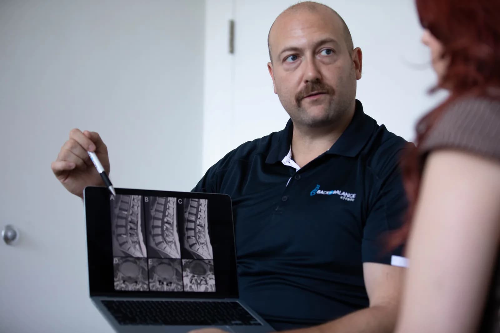
MRI provides much more detailed images of discs, nerves, the spinal canal and other soft tissues.
When advanced imaging of the lumbar spine is clinically indicated, MRI is often the preferred test, particularly when we are concerned about neurological compression, cancer, infection or when imaging is needed to help plan certain spinal procedures.
MRI also has the advantage of not using ionizing radiation.
CT Scan
CT provides excellent detail of bone and may be appropriate in particular circumstances, including some types of trauma or when detailed assessment of bony anatomy is required.
It can also sometimes be used when MRI is indicated but cannot be performed.
Unlike MRI, CT uses ionizing radiation.
Why Isn't My Low Back Pain Showing Up on My MRI?
This can be frustrating.
Someone can have significant back pain and still have an MRI that doesn't identify a clear structural explanation.
That does not mean the pain isn't real.
Pain is more complicated than a picture of anatomy.
An MRI is very good at showing certain structures, but it doesn't directly measure pain. It also doesn't show us everything about how your spine behaves when you move, how sensitive a particular structure may be, your strength or physical capacity, or the many biological and contextual factors that influence pain.
This is one reason a thorough history and physical examination remain so important.
The opposite situation is also common: someone can have substantial changes on an MRI and very little or no pain.
The relationship between structure and symptoms is real — but it isn't one-to-one.
When Is Imaging Indicated for Low Back Pain?
There are situations where imaging is appropriate and sometimes urgently necessary.
The decision depends on your history, examination and what condition your healthcare provider suspects.
Examples include concern for:
- fracture, particularly following significant trauma or in someone at increased fracture risk
- cancer
- spinal infection
- cauda equina syndrome
- severe or progressive neurological deficits
- epidural abscess or hematoma
Imaging may also become appropriate when persistent or progressive symptoms have not responded as expected and the result would help guide the next stage of management — particularly if a specialist procedure or surgery is being considered.
Importantly, a single “red flag” does not automatically mean that someone has a serious condition.
For example, older age by itself is not a particularly accurate screening test for spinal malignancy. A previous history of cancer is considerably more informative.
Clinicians therefore need to interpret risk factors together with the patient's symptoms and examination rather than treating every individual red flag as an automatic indication for an X-ray or MRI.
What About Sciatica or a Disc Herniation?
Having sciatica doesn't automatically mean you need an MRI either.
Symptoms from irritation or compression of a lumbar nerve can often be identified clinically based on the history and neurological examination.
In the absence of concerning features, immediate imaging is generally not recommended for uncomplicated low back pain with sciatica.
If leg symptoms are severe or progressively worsening, there is significant neurological loss, or symptoms persist despite appropriate management and an injection or surgical opinion is being considered, MRI may become much more useful.
At that point, the imaging has a purpose: it can help guide a treatment decision.
What If My Back Pain Has Lasted Longer Than Six Weeks?
This is another area where there is sometimes confusion.
You may have heard that everyone should get imaging once back pain has lasted four or six weeks.
That's not quite right.
Duration alone isn't necessarily an indication for imaging.
If your symptoms are persisting, the more useful first step is often a reassessment.
We want to know:
- Are your symptoms changing?
- Are they improving at all?
- Has your neurological examination changed?
- Is the original diagnosis still the most likely explanation?
- Have you had an appropriate trial of treatment and activity?
- Is there now reason to suspect a condition that requires investigation?
- Would an MRI or X-ray actually change what we do next?
If the answer to that last question is no, taking an image simply because a certain number of weeks has passed may not provide much value. This is why it is important to get assessed by someone with advanced training in spine care.
Can Getting an MRI or X-Ray Actually Be Harmful?
Imaging is generally safe when used appropriately, but unnecessary imaging has potential downsides.
Radiation exposure
X-rays and CT scans use ionizing radiation. MRI does not.
We shouldn't be frightened of medically necessary radiation, but we also shouldn't expose patients to it when the test is unlikely to provide useful information.
Incidental findings
The more we look, the more likely we are to find something.
An incidental finding can lead to additional imaging, referrals or procedures that may not ultimately help the patient.
Fear and the nocebo effect
The language surrounding imaging findings can also matter.
Being told you have a “degenerating spine,” “worn-out discs” or multiple abnormalities can understandably change how you view your body.
Research into diagnostic labels in musculoskeletal pain suggests that more specific structural labels may increase perceived seriousness and preferences for imaging or invasive treatment.
This doesn't mean we should hide imaging findings from patients. Quite the opposite.
We need to explain what they mean.
A disc bulge may be relevant to your symptoms. It may also be an incidental finding that has been present for years without causing a problem.
Context matters.
Healthcare resources
Imaging also uses healthcare resources.
When a test isn't clinically indicated, avoiding it means those resources remain available for people who actually need diagnostic imaging.
This is particularly relevant in a publicly funded healthcare system such as Ontario's.
If I Don't Get Imaging, How Do You Know What's Wrong?
This is probably the most important question.
Not ordering imaging is not the same thing as not investigating your back pain.
A good low back assessment starts with a detailed history and physical examination.
Depending on your presentation, that can include assessing:
- how your symptoms started
- where your pain is located and whether it travels
- movements and activities that change your symptoms
- strength
- sensation
- reflexes
- nerve tension
- movement and functional capacity
- relevant medical history
- risk factors for serious pathology
The purpose is first to determine whether your presentation is consistent with a musculoskeletal problem that can be managed conservatively or whether further investigation or referral is appropriate.
Sometimes the result of that assessment is: we should get imaging.
Often, it isn't.
Both can be appropriate clinical decisions.
When Should I Seek Urgent Assessment for Low Back Pain?
Most low back pain isn't an emergency, but some symptoms require prompt medical assessment.
Seek urgent medical attention if back pain is associated with symptoms such as:
- new difficulty controlling your bladder or bowels
- difficulty initiating urination or new urinary retention
- numbness around the groin, genitals or saddle region
- severe or rapidly progressive weakness in one or both legs
- significant trauma with concern for spinal injury
- fever or systemic illness with significant back pain, particularly when infection risk is present
A history of cancer, unexplained systemic symptoms, significant fracture risk or progressive neurological changes should also be discussed promptly with an appropriate healthcare provider.
So, Should I Get Imaging for My Low Back Pain?
For most people with a new episode of uncomplicated low back pain, probably not.
Current evidence and clinical guidelines consistently recommend against routine imaging when there are no signs of serious pathology and when the result isn't likely to change treatment.
That doesn't mean imaging is bad.
It means imaging is most valuable when we're asking it a specific question.
An X-ray, CT or MRI should be one piece of the clinical puzzle — not a substitute for taking a good history, performing an appropriate examination and understanding the person experiencing the pain.
If you're unsure whether your back pain needs imaging, an appropriate next step is a clinical assessment. We can determine whether your presentation is suitable for conservative management or whether imaging, medical assessment or specialist referral is warranted.
References
Brinjikji W, Luetmer PH, Comstock B, et al. Systematic literature review of imaging features of spinal degeneration in asymptomatic populations. AJNR American Journal of Neuroradiology. 2015;36(4):811–816. doi:10.3174/ajnr.A4173.
Chou R, Fu R, Carrino JA, Deyo RA. Imaging strategies for low-back pain: systematic review and meta-analysis. The Lancet. 2009;373(9662):463–472. doi:10.1016/S0140-6736(09)60172-0.
Lancaster B, Goldman J, Kobayashi Y, Gottschalk AW. When is imaging appropriate for a patient with low back pain? Ochsner Journal. 2020;20(3):248–249. doi:10.31486/toj.20.0077.
Downie A, Williams CM, Henschke N, et al. Red flags to screen for malignancy and fracture in patients with low back pain: systematic review. BMJ. 2013;347:f7095. doi:10.1136/bmj.f7095.
American College of Radiology. ACR Appropriateness Criteria: Low Back Pain. American College of Radiology.
Choosing Wisely Canada. Radiology Recommendations: Don't do imaging for lower-back pain unless red flags are present. Canadian Association of Radiologists.
Choosing Wisely Canada. Spine Recommendations: Don't routinely image patients with low back pain unless serious underlying pathology is suspected or imaging is required to guide an evidence-based intervention.
National Institute for Health and Care Excellence. Low back pain and sciatica in over 16s: assessment and management (NG59). NICE.
Plinsinga ML, et al. The effect of diagnostic labels on treatment preferences and beliefs in people with musculoskeletal pain: a systematic review of randomized trials. Journal of Orthopaedic & Sports Physical Therapy. 2026;56(1):4–15. doi:10.2519/jospt.2025.13759.