
Most people do not need an MRI for low back pain when symptoms first begin. An MRI becomes useful when there is concern about a serious condition, severe or worsening nerve dysfunction, or persistent symptoms for which the results of the MRI change the proposed treatment plan (rehabilitation vs. surgery).
Wanting a scan is understandable. Your back hurts, it may be interfering with sleep or work, and you want to know what is happening. A detailed picture of what is going on seems like a sensible place to start.
Unfortunately, finding something on an MRI scan and finding something useful that changes your care are two different things. Before ordering an MRI, the useful question is: What would we do differently because of the result? NICE's imaging guidance follows that principle.
What can a lumbar MRI actually tell you?
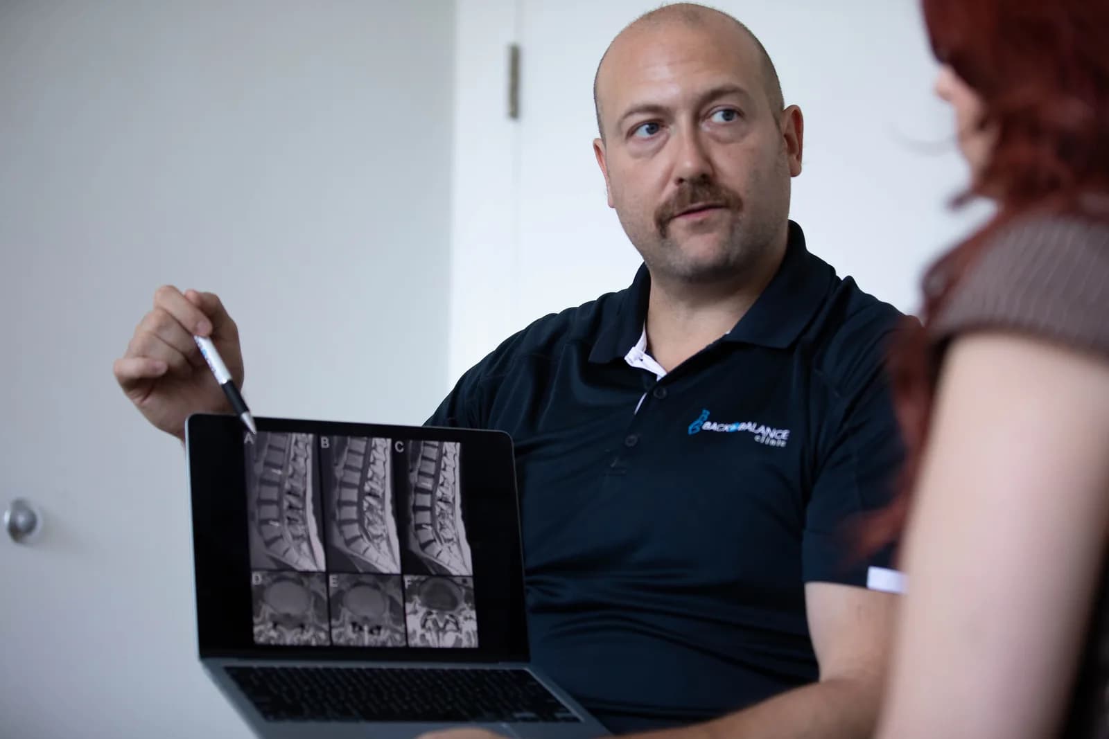
A lumbar MRI provides detailed images of the lower spine. MRI stands for magnetic resonance imaging; it uses a magnetic field and radio waves to show structures such as discs, nerves and surrounding tissues. Unlike x-rays and CT scans, MRI does not use ionizing radiation.
MRIs are very good at seeing soft tissues (muscles, discs and nerves), and therefore can help identify nerve compression, disc herniations, infections, tumours and other conditions. What it cannot reliably do is identify the source of every person's back pain from the image alone.
For example, a disc protrusion affecting a nerve on the same side as your leg symptoms may be relevant. However, a small bulge elsewhere may have little to do with why sitting hurts this week. The scan needs to fit the history and clinical examination. Everyone is likely going have some structural changes on MRI that become more frequent as we get older, but that does not mean that each of those findings is the cause of this current low back pain episode.
This has lead to the use of the term nonspecific low back pain. It means that a single, definite cause has not been established and no specific serious condition has been identified. It does not mean the pain is imaginary or that an assessment has found nothing useful. It just means there is not a clear structural cause of low back pain.
Why an MRI for low back pain is often unnecessary
Studies show that routine early imaging does not improve pain or function in people without signs of a serious underlying condition. That is the main reason guidelines advise against scanning everyone with a sore back.
A 2026 systematic review showed that compared to treatment that did not include immediate imaging, getting a lumbar MRI before starting care did not improve pain or functional outcomes. Importantly, this evidence is most applicable to recent, uncomplicated back pain; it should not be used to dismiss someone with a concerning examination.
Consider someone whose back became painful after gardening. Their neurological examination is reassuring, there are no concerning medical features, and they are gradually improving. Whether or not you confirm the clinical diagnosis with an MRI, the initial plan may still involve staying active within tolerance, temporarily adjusting aggravating tasks and progressively rebuilding activity.
An MRI can confirm a label without improving that plan. If we think it is a disc bulge, and the MRI shows a disc bulge, the treatment plan is the same. The only thing that changed is many patients may delay care waiting on the MRI results, and then are behind where they could have been functionally if they had just started their rehabilitation plan immediately.
What symptoms make imaging urgent?
Some symptoms require urgent medical assessment, with MRI or other investigations arranged according to the suspected problem. These warning signs are often called red flags.
New bladder, bowel or saddle-area symptoms
With back or leg pain, new difficulty starting urination, inability to empty the bladder, loss of awareness of bladder filling, new urinary leakage, loss of bowel control, or numbness around the genitals, anus or inner thighs warrants emergency assessment. Severe or rapidly worsening leg weakness is also concerning.
These can indicate cauda equina syndrome, a condition involving compression of the bundle of nerves at the bottom of the spinal canal. It can affect bladder, bowel and leg function. Do not wait for several warning signs to appear or for a routine outpatient scan.
Possible cancer, infection or fracture
Prompt medical assessment is also needed when back pain occurs in a concerning context, such as:
- A history of cancer with new, unexplained back pain, particularly alongside other concerning symptoms.
- Fever or feeling systemically unwell, especially with a recent infection, spinal procedure, immune suppression or injection drug use.
- Significant trauma, or new pain after a relatively minor event in someone with osteoporosis or prolonged corticosteroid use.
Someone who is very unwell or has neurological changes should seek emergency care. MRI is often useful for suspected infection or cancer; a suspected fracture may initially require an X-ray or CT. The best test depends on the question.
While the presence of a red flag raises concern, it is not a diagnosis in isolation. Many individual warning signs have limited accuracy, so clinicians consider the whole presentation and clusters of symptoms rather than any one symptom alone. Night pain alone, for example, does not automatically mean cancer or require an MRI. Equally, a concerning history should not be brushed aside because one symptom is absent.
Does sciatica or persistent symptoms justify an MRI?
Sometimes, but not automatically. It depends on the severity and whether or not your specific condition is a good candidate for surgery. Sciatica refers to pain travelling into the leg, originating in your sciatic nerve, commonly associated with irritation of a nerve root in the lumbar spine. However, leg pain does not automatically require immediate imaging.
If symptoms are stable, there is no significant or worsening weakness, and an invasive treatment is not being considered, an initial period of non-operative care is often indicated.
For persistent, disabling leg symptoms despite appropriate care, or progressively worsening neurological symptoms in the leg, an MRI can help determine whether there is a finding that matches the symptoms and could be treated surgically or with a selected injection.
For example, persistent right-sided leg pain with matching examination findings may justify imaging when a specialist is considering a procedure. The MRI helps answer a specific question: is there nerve compression at the expected level, and would surgically treating it help?
Does the six-week mark automatically mean you need a scan?
No. Around six weeks is a common point to reassess your progress and reconsider imaging in uncomplicated cases that have not improved. It is not an automatic MRI deadline. There may be other factors that are slowing healing, and if surgery is unlikely to address these factors, it may warrant changing the clinical approach vs. turning to imaging.
Guidelines differ somewhat for persistent pain confined to the back. The Canadian Association of Radiologists recommends an initial X-ray after unsuccessful conservative treatment, while NICE keeps imaging tied to whether it is likely to change management. Neither makes persistent pain alone an automatic indication for lumbar MRI. It is a decision that is going to be made by your clinician based on the overall clinical picture.
Ongoing pain deserves reassessment. That may reveal a reason to investigate, a need to revise treatment, or both.
Does a disc bulge on MRI explain your pain?
It might, but its presence alone does not prove that it is causing your symptoms.
Common spinal changes also occur in people who have no back pain.
A systematic review of 3,110 people without symptoms estimated the following age-related prevalence:
Disc degeneration:
- Age 20: 37%
- Age 40: 68%
- Age 60: 88%
Disc bulge:
- Age 20: 30%
- Age 40: 50%
- Age 60: 69%
Disc degeneration describes changes in disc structure, including water content and height. Disc bulge refers to a change in the circumference of a disc. These diagnoses do not, by itself, establish how painful or capable your back is.
There is another side to this evidence. Some findings are more common in people with back pain than in those without it. It would be inaccurate to say all degeneration is irrelevant. However, the finding's location, severity and relationship to your symptoms matter. A report needs clinical interpretation, rather than a blanket statement that everything is either damaged or normal. The imaging findings may be a useful way to confirm clinical findings, or differentiate between two potential conditions, but it should not be assumed that everything seen on imaging is automatically clinically relevant.
Can unnecessary imaging make back pain worse?
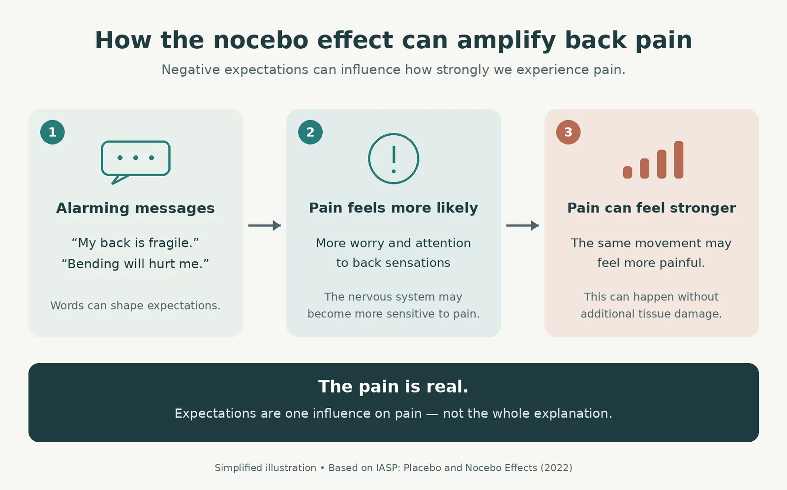
It can contribute to a harder recovery in some people, but it is very person specific.
One main area of concern is the nocebo effect: a psychobiological phenomenon where a person experiences real physical side effects or worsening symptoms, because they expect a treatment or condition to be harmful, even if it is completely inert or harmless.
Terms such as “degeneration” or “disc disease” may lead someone to believe their spine is fragile. That belief can influence how they experience pain and approach movement. Negative expectations can affect pain processing and the resulting pain is real.
In practical terms, someone who was beginning to walk comfortably may stop exercising after reading a report, fearing that every bend is causing further "damage". Reduced activity and growing fear may then make returning to normal life more difficult in some people.
A small randomized study of 44 people with low back pain found that those given a conventional explanation of their MRI report had less improvement in pain and poorer functional outcomes than those reassured that the findings represented normal changes. This supports the idea that how results are communicated are just as important as the findings themselves. For patients at Back in Balance, we like to walk patients through their imaging findings and explain them in plain English so they can understand what may be relevant to their condition, and what are incidental findings.
So- What should happen before you decide on imaging?
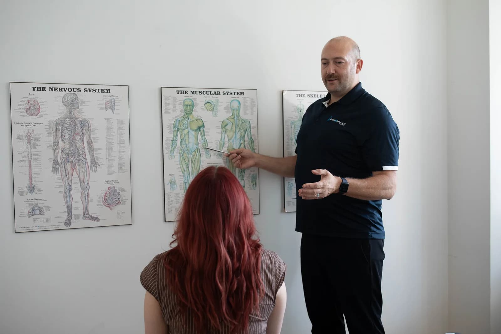
An assessment should establish what your symptoms are doing, examine nerve function where appropriate, consider your medical history and explain the next step. If imaging is recommended, you should understand what the clinician is looking for and how the result might change care.
If imaging is deferred, there should still be a plan: what to do now, when to reassess, and which changes should prompt earlier medical attention. You should not have to choose between an unnecessary scan and being told to simply put up with the pain.
At Back in Balance Clinic in Toronto, an initial assessment can help determine whether your low back pain needs imaging, a treatment plan, or medical referral. Emergency warning signs require emergency care.
Book an initial assessment at Back in Balance Clinic.
Useful sources
- NICE: Low back pain and sciatica in over 16s, NG59, updated 2020. Imaging is considered when it is likely to change management.
- Canadian Association of Radiologists Spine Imaging Referral Guideline, 2025; first published online in 2024. Canadian recommendations for routine, urgent and condition-specific imaging.
- Choosing Wisely Canada: Radiology recommendations, updated June 2026. Practical Canadian guidance on avoiding unnecessary back imaging.
- American College of Radiology: Low Back Pain Appropriateness Criteria. Recommendations include MRI for selected intervention candidates with persistent symptoms after approximately six weeks of care.
- Skelly and colleagues: Assessment and Treatment of Acute Low Back Pain: A Systematic Review, 2026. Recent synthesis finding no improvement in pain or function from early imaging without signs of serious spinal pathology.
- Brinjikji and colleagues, 2015: spinal imaging findings in people without symptoms and comparison with symptomatic adults. Read together to understand both common incidental findings and genuine associations with pain.
- Rajasekaran and colleagues: The catastrophization effects of an MRI report, 2021. Small randomized trial examining the effect of how MRI findings are explained.
- Characteristics and Effectiveness of Interventions That Target the Reporting, Communication, or Clinical Interpretation of Lumbar Imaging Findings: A Systematic Review, 2022. Important limitations in the evidence on imaging communication.
- Lemmers and colleagues: Imaging versus no imaging for low back pain, 2019. Systematic review of costs, healthcare use and work absence.
- Downie and colleagues: Red flags to screen for malignancy and fracture, 2013. Foundational systematic review showing the limitations of individual warning signs.